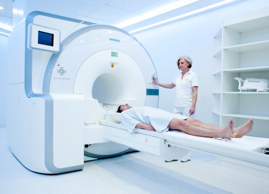
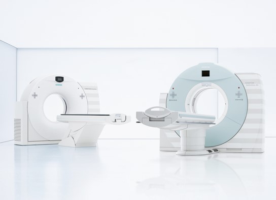
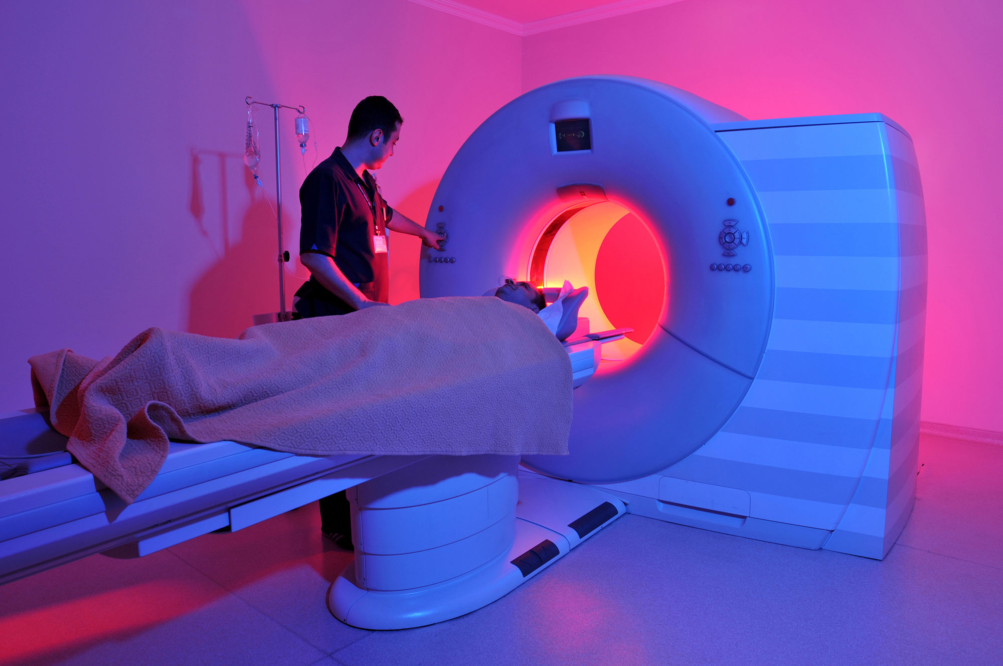
The Hospital operates an ultramodern Nuclear Medicine Department, equipped with state-of-the-art technology. Both in vivo diagnostic tests and the therapeutic application of radioisotopes are performed at the Nuclear Medicine Department.
IN VIVO DIAGNOSTIC TESTS
In vivo diagnostic tests are performed with all the necessary radiopharmaceuticals and routine techniques. The following test are performed: scintigraphies of the thyroid gland (using Tc-99m, I-131 or Tl-201), bones (plain - three phase), kidneys (static – dynamic renal scintigraphy), lungs, sentinel lymph node, gastric emptying time, lacrimal duct, gastrointestinal bleeding, brain (perfusion-carotids, tomography, statics, etc.), parathyroid glands, heart (static/dynamic, stress and resting ventriculograph, first pass for communications, etc.), liver, biliary paths, gallbladder, spleen, saliva gland, etc., whole body scintigraphies (using Ga-67, Tl-201 or I-131).
TREATMENTS USING RADIOISOTOPES
- Radioactive iodine I-131 therapy for thyroid cancer (average inpatient stay of 2 days in a room that is specially designed for radioisotope treatment)
- Radioactive iodine I-131 therapy for hyperthyroidism (average inpatient stay of 2 days in a room that is specially designed for radioisotope treatment)
- Radioactive iodine I-131 therapy for goiter (average inpatient stay of 1 day in a room that is specially designed for radioisotope treatment)
- Palliative treatment of bone metastasis from prostate/breast cancer with radiopharmaceuticals such as strontium, rhenium, samarium, etc. (average inpatient stay of 2 days in a room that is specially designed for radioisotope treatment)
- Treatment of arthritis, radiosynoviorthesis, radiosynovectomy and radiation synovectomy using rhenium, erbium, yttrium, etc. (average inpatient stay of 2 days in a room that is specially designed for radioisotope treatment)
- Treatment with In-111 Octreoscan for somatostatin receptor-positive tumors
- Treatment of pheochromocytoma, neuroblastoma, thyroid gland carcinoid and medullary tumors with I-131 ΜΙΒG
PET/CT
A high-tech imaging method combining Positron Emission Tomography (PET) and Computed Tomography (CT).
It brings about remarkable changes in the management of cancer patients.

What are the applications of PET/CT?
- Maps the extent of the disease (staging)
- Checks response to treatment
- Promptly detects recurrence (restaging)
- Offers radiotherapy planning
- Monitors the course of the disease
- Detects disease in case of elevated cancer markers
- Offers biopsy guidance

These apply to the majority of cancers such as: lung, Hodgkin and Non-Hodgkin lymphoma, breast, esophagus/stomach, melanomas, large intestine, head/neck, gynecologic cancers, sarcomas, cancers of unknown primary site, seminomas.


