


The Center is equipped with two state-of-the-art digital mammography systems Hologic – Selenia, one of which offers advanced three-dimensional imaging (Tomosynthesis). It also features a high-resolution ultrasound system, specifically designed for breast examinations. In the same center, a stereotactic biopsy system (BLESS) is installed, allowing for highly precise and minimally invasive diagnostic procedures.
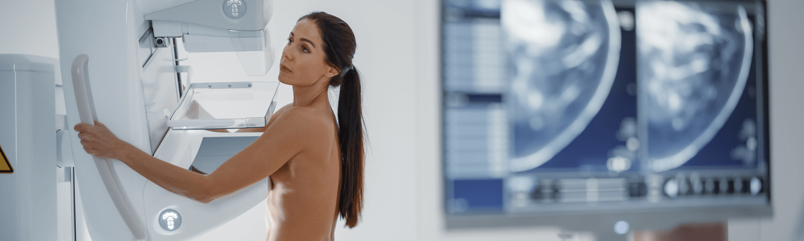
1. Digital Mammography
Comprehensive breast imaging is achieved through mammography, which enables the assessment of the health of both breasts. The use of advanced digital technology provides images with greater contrast and enhanced detail, a factor of critical importance in the early detection of abnormalities.
Mammography remains the only globally accepted method for breast cancer screening. In many European countries—though not yet in Greece—it has been incorporated into national population-wide preventive health programs.
Experience from such large-scale screening initiatives has shown a measurable reduction in both morbidity and mortality among women, while also generating significant social and economic benefits for the countries that adopt them. Unfortunately, in Greece, the development of such wide-reaching preventive strategies is still at a very early stage.
2. Tomosynthesis
When deemed necessary based on the results of standard digital mammography, tomosynthesis is performed as a complementary technique. This advanced method, often referred to as 3D mammography, represents the evolution of digital mammography, offering three-dimensional imaging of the breast. It enables earlier, more accurate, and more detailed diagnoses compared to traditional 2D mammography. Using 1 mm slice acquisitions, images are reconstructed, allowing the detection of lesions that may not be visible with standard mammography. This significantly reduces the likelihood of false-negative results.
Tomosynthesis is particularly valuable for women with dense breast tissue, where conventional methods have often made timely and accurate diagnosis more challenging.
Detecting malignancies at an early stage is of paramount importance, as it improves survival rates and enhances prognosis.
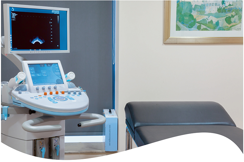
3. High-Resolution Ultrasound – Elastography
While mammography remains the primary imaging method for breast cancer screening, it has certain inherent limitations in detecting malignancies at an early stage. These limitations are especially evident in women with dense breast tissue—more common among younger women—or when specific tumor subtypes mimic the appearance of normal tissue.
This well-documented constraint increases the likelihood of false-negative results. To reduce this risk, high-resolution ultrasound is often incorporated into routine screening, as it can reveal lesions that mammography alone may overlook.
At our center, high-resolution ultrasound is performed by highly experienced radiologists using state-of-the-art imaging equipment. This approach allows for the detection of very small lesions (less than 5 mm), which are usually identified at an early stage.
In addition, Shear Wave Elastography is applied alongside ultrasound in selected cases to further enhance diagnostic accuracy. This method assesses the stiffness or elasticity of a lesion—since malignant tissue is typically much firmer—by quantifying it in kilopascals (kPa). This provides a reliable distinction between benign and suspicious findings.
When required, ultrasound-guided biopsies are also performed. This globally accepted gold-standard method not only confirms diagnosis but is also an essential step in preoperative planning for malignant cases. The procedure is minimally invasive, completed within minutes under local anesthesia, and carries no complications, ensuring both safety and diagnostic precision.
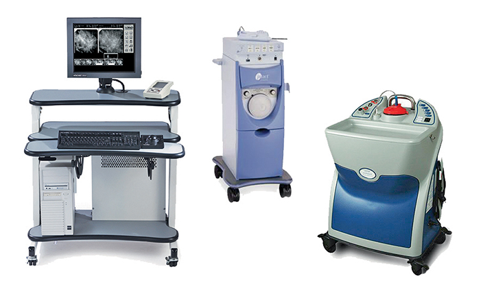
4. Stereotactic Biopsy
Stereotactic biopsy is an advanced, minimally invasive technique for diagnosing suspicious breast lesions, helping to avoid unnecessary surgical procedures and ensuring the patient’s safety and comfort.
It is most often recommended when mammographic findings—such as clusters of calcium deposits (microcalcifications)—are considered suspicious, as these may sometimes be linked to malignant breast disease.
The procedure is simple and almost fully automated, performed with specialized equipment that guarantees high precision and accuracy, thus ensuring a successful surgical procedure.
5. Lesion Localization with Wire Guidance
When a non-palpable breast lesion, visible only through imaging, requires surgical removal, a fine wire guide is placed preoperatively. This enables the surgeon to precisely locate the lesion within the breast, making excision easier, safer, and more accurate.
Placement of the wire is performed with utmost precision using either 3D tomosynthesis or high-resolution ultrasound, ensuring maximum accuracy and patient comfort.
Magnetic Resonance Imaging – Advanced Imaging: Breast