Metropolitan Hospital operates a model, fully equipped and organized Urology Clinic. Staffed by qualified scientific and nursing personnel and equipped with state-of-the-art technology, the Urology Clinic is open to patients and ready to address any acute and/or chronic disorder of the genitourinary system at any time.
Fully qualified and trained, the medical and nursing staff offer our patients all the diagnostic, therapeutic and surgical services, thereby achieving ideal assistance and inpatient support.
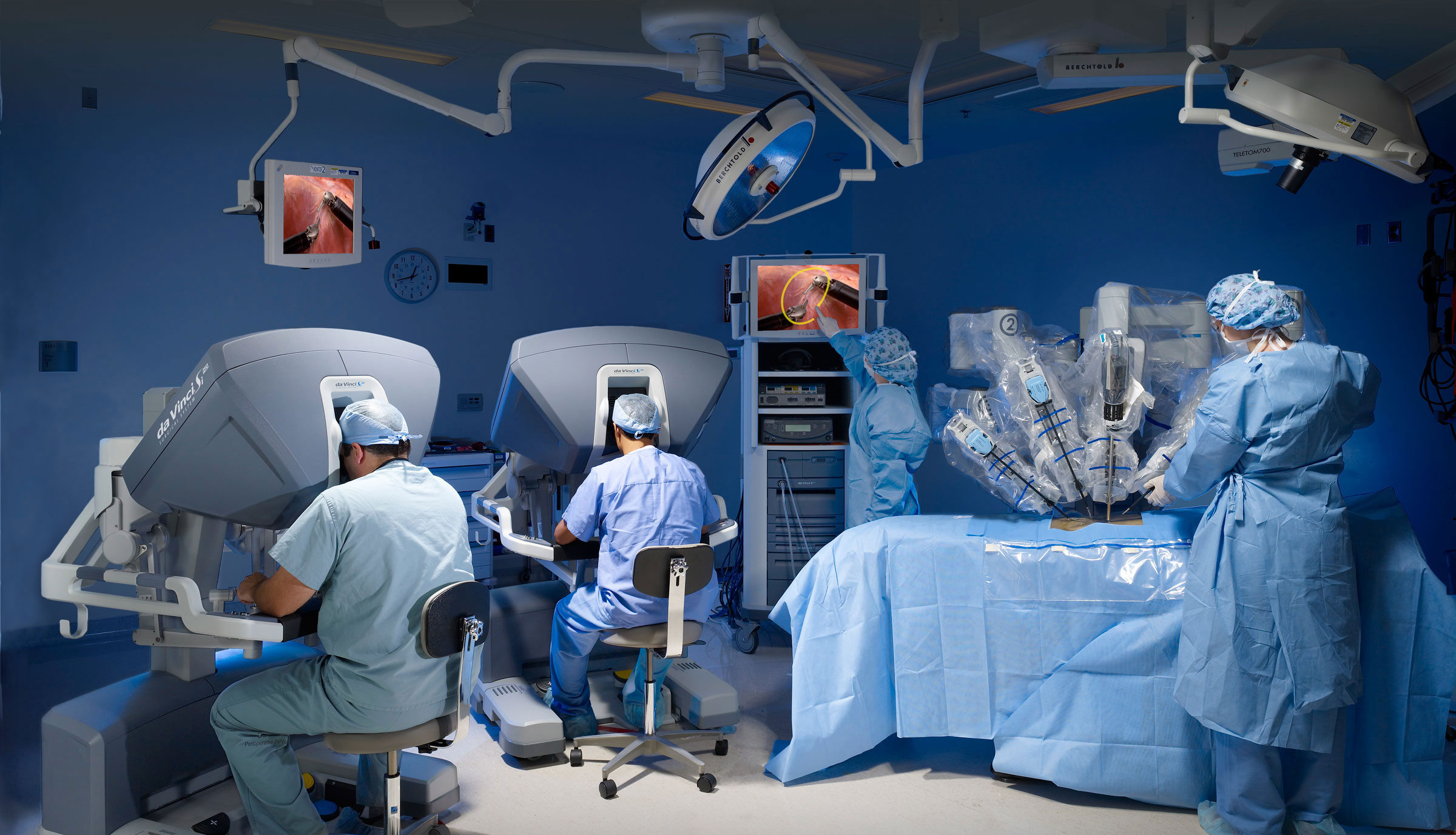
The da Vinci Si, a high-tech robotics system, assists in the treatment of all urologic oncology and benign disorders, by performing minimally invasive, but difficult procedures, such as radical prostatectomy with lymphadenectomy, radical cystectomy with neobladder, radical or partial nephrectomy or nephroureterectomy.
SURGICAL CHOICES
The therapeutic choices for urologic conditions may include one or more of the following and depend of the stage of the disease and the patient’s overall health:
- Radiation therapy
- Biological therapy
- Open surgery
- Chemotherapy
- Minimally invasive surgery
Open surgery
In the case of open surgery, a large incision is made in the abdomen. The exact size of the incision depends on the type of procedure that will be performed. Open surgery, also known as laparotomy, allows the surgeon to touch the organs and feel the inside of the patient’s body.
Minimally Invasive Surgery / Laparoscopy
The surgeon stands next to the patient and operates via small openings, using long, straight instruments and a microscope camera.
Robotically-assisted surgery
The three-dimensional, high resolution camera provides a magnification of the inside of the patient. The camera sends images to a monitor in the operating room to guide surgeons during the procedure. The surgeon sits at a console and controls the instruments, which bend and rotate like two tiny hands on the inside of the patient. The surgeon uses a three-dimensional, high-resolution system, which provides a magnification of the inside of the patient. The system converts all hand movements to smaller, precise movements of the tiny instruments on the inside of the patient.
ROBOTIC UROLOGY (DA VINCI)
Nowadays, robotic prostate cancer surgery is performed on 85% of case in the US. Being at the forefront of innovations, Metropolitan Hospital is not only equipped with the latest state-of-the-art da Vinci system, the da Vinci Si, but also has one of the most experienced and qualified teams of Greek robotic surgeons, who perform the largest number of procedures in Europe annually.
READ MOREONCOLOGY
Oncology addresses the entire urologic oncology spectrum and performs significant surgical procedures, such as radical prostatectomy (accessed laparoscopically), radical cystectomy and ileectomy or neobladder, and lymph node removal.
Today, the latest brachytherapy method for prostate cancer (real-time brachytherapy) is performed at Metropolitan Hospital.
The Oncology Department is supported by the Oncology Council, which examines the diagnoses and data that arise from tests, and decides and recommends the best and most effective treatment.
See also
Advanced MRI: Prostate
NEPHROLITHIASIS CLINIC
What is urinary lithiasis
It is defined as the presence of one or more stones in any part of the urinary tract. Stones may be found in the kidneys, the ureters (the tubes that transfer urine from the kidneys to the urinary bladder), the urinary bladder itself or the urethra.
The size of the stones varies from a few millimeters to quite a few centimeters. Some stones occupy the entire renal collecting system, the pelvis and the calyces, forming a casting of the cup-and-pelvis system and are known as coral stones.
Why stones are formed
Kidney stone formation is due to the increased concentration of certain substances in the urine, allowing the crystallization of these substances and the increased size of the stone. Increased concentration may either be due to a reduced urine volume due, possibly to low fluid intake, or the increased elimination of these fluids in the urine due to medical conditions.
Depending on the substances the stones are made of, they are mainly classified into:
- Calcium oxalate and calcium-phosphate stones, which are the most common
- Cystine stones: individuals suffering from cystinuria
- struvite/infection stones, in individuals suffering from chronic urinary tract infections, mainly women.
- Uric acid stones, which are hard stones, usually not depicted in simple X-rays. They are common in chemotherapy patients, mainly men.
Predisposing factors
Age/Gender: Nephrolithiasis is most common in men in the 20-60 age group.
Inheritance: Almost 25% of nephrolithiasis patients report a family history of stones.
Medical conditions: The possibility of kidney stone formation is heightened in any condition in which there is a reduced volume of urine or increased elimination of various substances in the urine that favor stone formation.
- Low fluid intake and dehydration.
- Congenital abnormalities of the urinary tract that may partially or completely obstruct the flow of urine.
- Obstructive diseases of the urinary tract such as prostatic hyperplasia.
- Elevation of substances in the blood and urine such as calcium (hypercalcemia), uric acid (hyperuricemia) and cystine (cystinuria).
- Thyroid disorders, especially of the parathyroid glands.
- Administration of calcium supplements e.g. for osteoporosis and chemotherapy drugs.
Dietary habits: Diet rich in calcium, oxalates, saturated fats, proteins, glucose and vitamin C raise the risk of forming kidney stones.
Nephrolithiasis and diet
Many discussions have been conducted on this issue, but without arriving at clear conclusions. The substances that participate in the formation of stones are an integral part of our diet and cannot be excluded from it. Studies agree that the body’s sufficient hydration reduces the risk, without ruling out kidney stone formation. Diets that are rich in protein, animal fat, sugar and salt appear to raise kidney stone formation risk. The diet that is recommended to each nephrolithiasis patient should be customized according to their needs and stone composition. In any event, a balanced diet in combination with physical exercise and maintenance of normal body weight are a common factor in treating nephrolithiasis.
Renal colic / What causes it
When a kidney stone of any size partially or completely obstructs the normal flow of urine, then the sudden increase in pressure within the urinary tract causes the characteristics and intense symptoms known as renal colic. This usually occurs when a stone travels to the ureter and gets lodged along the way. The obstruction in turn causes the swelling of the other kidney-ureter; this is known as hydronephrosis.
Symptoms of nephrolithiasis
- Intense pain in the kidney area that fluctuates, with referred pain in the corresponding abdominal area down to the external genital organs.
- Sweating, nausea and vomit.
- Frequent urination and a feeling of being unable to urinate.
- Hematuria.
- Fever and physical discomfort.
- Interrupted urination in the case of bladder stones.
Renal colic is usually a severe condition in which the patient seeks medical assistance!
On some occasions, nephrolithiasis DOES NOT present the characteristic symptoms of colic, but rather some disturbances in the kidney area at sparse intervals, which the patients do not pay particular attention to. In some cases the stone needs to grow significantly before giving any symptoms. This is the reason why any disturbance in the kidney region, whether mild or intense, must be investigated by a urologist.
What should I do when I’m in pain
Nephrolithiasis is a very common disorder among both men and women. Many people have someone in their environment who has experienced colic. This, in combination with specific pain, leads many to making their own diagnosis and empirically treating the pain with painkillers, without ever consulting a doctor. Naturally, this is wrong because if innocent nephrolithiasis is underestimated, it can lead to serious conditions, with severe consequences. In recent years, many studies have shown that immediate and prompt treatment of nephrolithiasis reduces the possibility of complications and morbidity by more than 50%. Therefore, every time that we experience one of these symptoms, we must immediately consult a urologist.
How kidney stones are diagnosed
- Medical history and clinical examination.
- Simple X-ray of the kidneys/ureters/bladder (KUB): It may not provide much information when the stone is small or radiotransparent
- KUB ultrasound: An ultrasound can diagnose 3mm or larger stones in the kidney or the upper part of the ureter. Even if the ultrasound is unable to detect a stone, we check for dilation, namely the result of obstruction.
- Abdominal CT: It is by far the examination of choice. It reveals stones of any size and at any location in the urinary system. It provides us with valuable information about the state and functionality of the kidney.
- Urine and blood tests: Patient is tested for the growth of a bacterial infection within the urinary system, which possibly accompanies nephrolithiasis and greatly affects clinical appearance.
Treatment
The importance of correct and prompt treatment has already been stressed:
- Small stones measuring up to 3mm, which do not cause significant obstruction and are not accompanied by an infection, are monitored pending their elimination.
- Larger, lodged stones that cause obstruction and are accompanied by infection must be surgically removed. The aim is to prevent permanent kidney damage and to reduce morbidity of the condition, which can develop into a life-threatening one.
The placement of a urinary stent (double-j or Pig Tail) along the length of the obstructed ureter is used to remove an obstruction. This is a fine stent that is inserted in the ureter under fluoroscopy guidance. The procedure is simply completed within 10 minutes, without the need for anesthesia. The obstruction of the kidney is removed, immediate risk is prevented and conditions are created for permanent treatment, which is ultimately to break the stone into smaller pieces.
Advanced, minimally invasive techniques
- Remote lithotripsy: The traditional method. This is a non-invasive method that is performed without anesthesia and the entire procedure lasts about 45 minutes.
- Laser lithotripsy: This is the most ideal and recent method with success rates that touch on 100%. It is endoscopically performed via the urethra and is bloodless. The patient is discharged within 24 hours.
- Percutaneous micro-lithotripsy: It is applied to large stones and corals. It is performed via a small 5mm incision in the skin, it is virtually bloodless and hospitalization lasts 24 hours.
The choice of lithotripsy method depends on a number of factors, mainly the size and position of the stone. Open surgery for nephrolithiasis is a thing of the past. Technological developments allow us to easily, quickly, non-invasively and safely treat any stone. The patient undergoes minimal stress and can return to their daily activities immediately. This, of course, requires that the surgeon is familiar with the advanced technology that is required to perform the delicate endoscopic procedures.
Nephrolithiasis Clinic
9 Ethnarchou Makariou & 1 Venizelou Streets, 18547 Neo Faliro
Wednesday, 12:00pm-1:30pm
Tel: +302104809325, +302104809150, +302104809160
ENDOSCOPY
Metropolitan Hospital boasts one of the most modern endoscopy departments in Greece today. In addressing urologic disorders, state-of-the-art technology has made it possible for the patient’s hospital stay to be reduced and surgical procedures are marked by greater safety and success.
GreenLightLaser is now used in the treatment of benign prostatic hyperplasia and lithiasis is performed endoscopically using the Holmium laser.
ANDROLOGY
Andrology addresses all disorders and cases related to male infertility and impotence, by performing all the latest diagnostic tests, such as penile ultrasound (triplex), cavernosography and semen analysis, as well as therapies for the treatment of erectile dysfunction and infertility.


