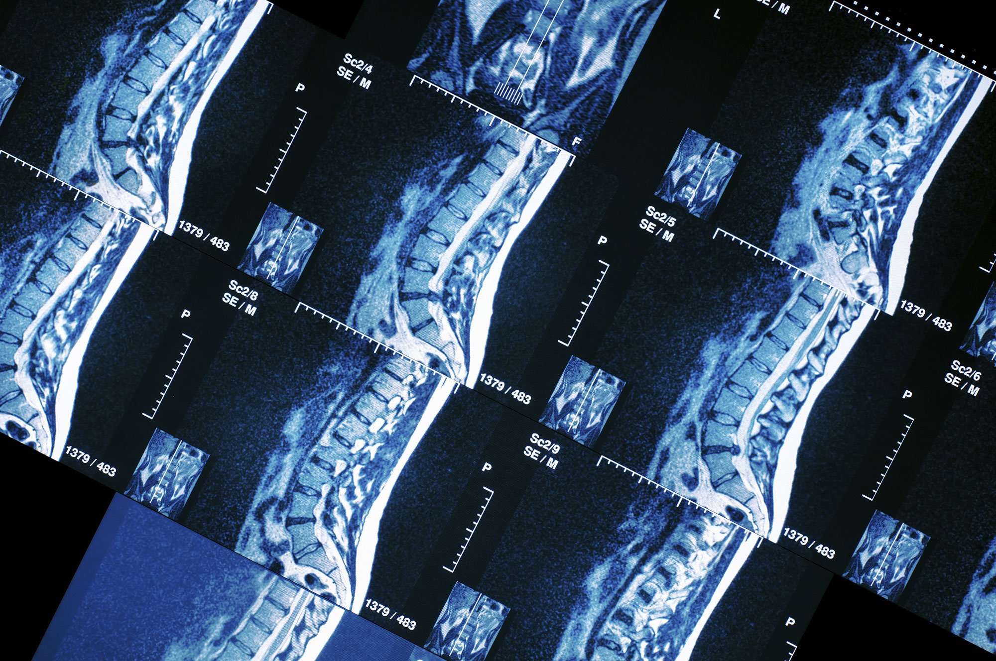
The special MRI techniques play a significant role in detecting and assessing the extent of clinically significant prostate cancer. These techniques significantly increase diagnostic accuracy when identifying and characterizing a lesion, permitting targeted biopsy collection from suspect areas (and not blindly, with the risk of not collecting material from the cancerous area). In addition, these special MRI techniques assist in estimating the extent of the disease (staging) with greater accuracy before surgery or radiotherapy. After treatment, they are used for early recurrence detection.

A multiparametric study is the combination of special MRI techniques..
Multiparametric MRI combines special techniques with high-resolution anatomical images, significantly increasing accuracy when detecting cancer.
At our Unit, multiparametric studies are performed using a state-of-the-art latest generation Siemens Skyra 3 Tesla scanner with a 60 channel dedicated abdomen coil, so the use of an endorectal coil is not necessary.
The protocol to be used and the interpretation of the results are determined by qualified doctors, based on the international guidelines of the American College of Radiology (ACR) and the European Society of Urogenital Radiology (ESUR) – PI-RADS Version 2 (2015).
See also
Robotic Urology (da Vinci): Oncology


