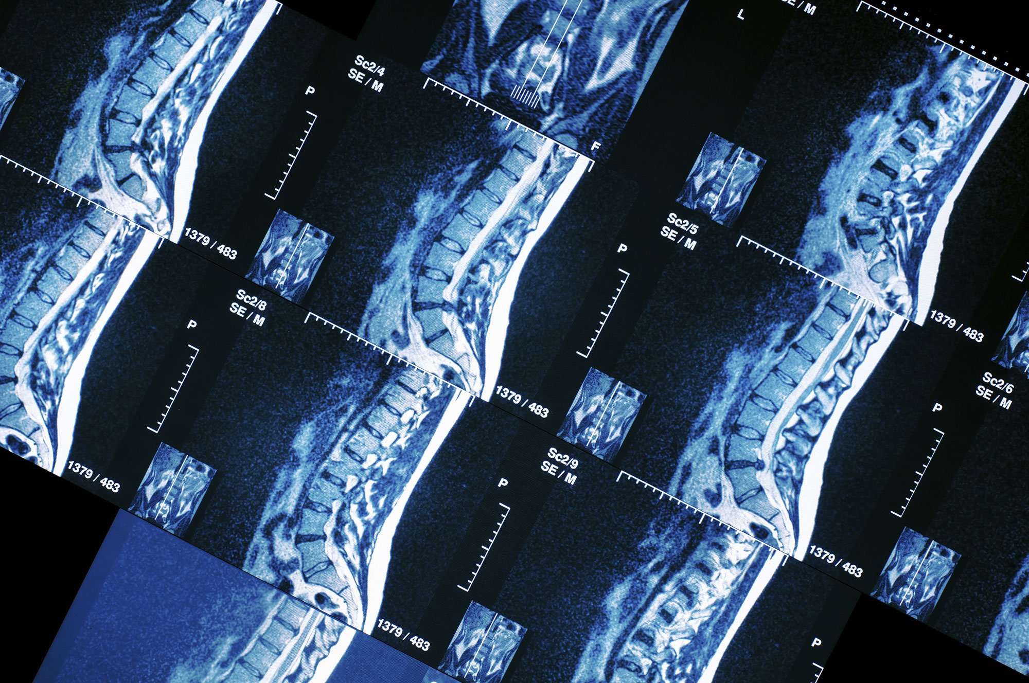
The special MRI techniques play a significant role in detecting and assessing the extent of breast cancer. These techniques significantly increase diagnostic accuracy when identifying and characterizing a lesion, contributing to selecting the most suitable treatment and to properly monitoring the treatment result.

ULTRA MODERN BREAST IMAGING
- With multiple applications in breast imaging (diagnosis, preoperative and radiotherapy planning, treatment result monitoring).
- With a team of qualified radiologists and radiophysicists, who apply special protocols in tune with international standards, adjusted to the anatomical area of the breast.
- With diagnosis offered by doctors with long experience who have received training in centers of excellence in the USA, have specialized in breast MRI and hold special licenses to this effect.
Breast MRI is a most significant tool in the early diagnosis and optimal treatment of breast cancer.
- It is very sensitive in detecting clinically significant cancer (invasive cancer and high-grade in situ carcinoma). It is superior to a mammography for dense breasts, as it may detect twice as many tumors.
- It is very important to apply the right technique, to process the data properly and to interpret the findings based on the American College of Radiology guidelines (ACR BI-RADS® – MRI Lexicon Classification Form).
- In our Unit, it is performed by qualified doctors*, with special multiparametric study techniques and post-processing of data in CAD (computer-aided diagnosis) systems.
- We are equipped with the latest technology and all the necessary components for breast imaging.
TECHNIQUES/EQUIPMENT
- Imaging using a special breast coil (18 channel dedicated breast coil) in a latest generation Siemens Skyra 3 Tesla scanner
- High-resolution images
- Special Diffusion techniques (multiple b value DWI, IVIM)
- Dynamic angiography (4D time resolved twist)
- Spectroscopy
- Data processing in CAD system
WHEN IS A BREAST MRI INDICATED?
The indications for a breast MRI keep expanding to include all the more women./p>
Complementary screening
It is indicated for women with a high risk of developing breast cancer, while in the last few years, it is increasingly being performed on women with a medium risk and women with dense breasts, using new, faster techniques (ultrafast breast MRI).
Solution to difficult diagnostic problems
It assists in characterizing a lesion in cases when the mammography and ultrasound findings are debatable. In many cases it may rule our cancer and women may avoid a biopsy or a surgical procedure. In other cases, it may confirm a malignancy and women may undergo the most suitable treatment right away.
Selection of the right treatment before surgery
It accurately identifies the extent of the cancer before surgery, so that surgeons select the most suitable treatment approach (lumpectomy or mastectomy, preoperative chemotherapy). Compared to mammography and ultrasounds, it estimates the size of the tumor with much greater accuracy. When the cancer has spread extensively, a woman may need to undergo a mastectomy or chemotherapy before the surgical procedure. On the contrary, if the tumor is small, the woman may undergo a lumpectomy and preserve the breast. Lastly, when the size of the tumor is accurately detected, total excision of the tumor may be achieved in one surgery, avoiding a second procedure (re-excision) in most cases.
Simultaneous assessment of the other breast (staging)
Monitoring of treatment result
It assesses the presence of residual cancer following a lumpectomy. For example, if the entire tumor was not removed, it accurately estimates the areas with residual foci (positive resection margins).
Cancer recurrence detection
It constitutes a prognostic indicator for response to neoadjuvant chemotherapy.
In these cases, it is indicated before, during and after treatment, preoperatively.
ULTRAFAST BREAST MRI
The new screening method:
- A specific group of women with relatively high risk of developing cancer may benefit from the new Ultrafast Breast MRI. This method is available at out Unit, in the context of screening tests, as a complementary method to a mammography. This group may include women with a positive medical or family history and women with dense breasts, given that a mammography may miss one in two lesions due to its very low sensitivity.
- An Ultrafast Breast MRI is a very fast and shorter version of the conventional MRI, lasting just 5 minutes. It is painless and does not expose women to radiation.
- Mammography screening may be complemented with other mammography techniques (tomosynthesis), with an ultrasound or with special ultrasonic techniques (elastography).
- A Breast MRI is more sensitive in identifying breast cancer, detecting at least double the number of lesions compared to a mammography and a much greater number even when combining a mammography with an ultrasound. Therefore, it is highly indicated as a screening test for high-risk women, according to international guidelines. However, the exam lasts about 15-20 minutes.
- Although an Ultrafast Breast MRI lasts just 5 minutes, studies have shown that it exhibits similar sensitivity to a conventional MRI in detecting lesions. It is recommended for women with dense breasts, given that a digital mammography is not as effective in detecting lesions. Therefore, it is a fast, highly sensitive and accurate method, which meets all the conditions for an ideal screening method. Distinguished doctors in the field of screening believe that it could play a significant part in breast screening for medium-risk women and women with dense breasts.
- In our Unit, it is recommended as a complementary exam to a digital mammography, wherever deemed necessary. However, it does not replace the digital mammography.
See also
Breast & Women's Center: Breast MRI
MRI-GUIDED BIOPSY
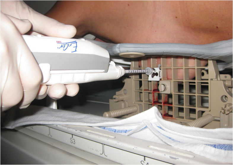
- The special MRI-guided breast biopsy technique available at Metropolitan Hospital is only performed in very few breast centers in Greece. This technique offers a unique way to collect a biopsy sample from a sub-category of breast lesions, which are only visible in an MRI. Such a biopsy is especially indicated for a suspect lesion that is depicted in a breast MRI, but is not visible during a digital mammography or an ultrasound.

- MRI-guided breast biopsy is performed by placing the woman in an MRI scanner in the prone position. Images are taken to accurately identify the location of the lesion. The biopsy sample is collected using local anesthesia on a small area of the skin. The procedure lasts about 1 hour and the woman may return home immediately.
- In our Unit, biopsies are performed by fully trained, highly skilled and suitably licensed doctors.


